


Francesca Galiano
|
31/08/2023 - Last update 14/11/2023
Yingzhi Li, Howe Liu, Charles Nichols, David C. Mason | Year 2022
Manual Therapy Treatment for Penile Pain- A Clinical Case Report with 6-Month Follow-up
Pathology:
Prostatitis and pelvic pain
Type of study:
Case Report
Date of publication of the study’:
2022/Apr/01
Purpose of the study
- Objective: to show the management of a case of penile pain through manual therapy, while highlighting the diagnostic and therapeutic clinical reasoning
- Measured outcomes: assessment of pain, range of motion (ROM) and Barthel Index to measure the level of autonomy in carrying out daily activities
Participants
- Number: 1
- Description: a 54-year-old farmer, father of 3 children with no history of genetic diseases, neither a smoker nor a drinker. No history of surgery, hospitalization or reproductive or sexual dysfunctions. A sharp and sudden onset of penile pain 3 weeks before, initially tolerable but then increased to an intensity of 9 out of 10 with daily dysuria after the first week. The pain occurred 4-5 times a night, for a duration of 10 minutes, and the only way to get some relief was to kneel forward in the fetal position while holding the penis in one hand.
In view of the urinary and intestinal problems resulting from the pain, the patient was hospitalized two weeks after the onset of the pain: although he could urinate or evacuate, he felt a lot of pain in the penis and in the genital areas. There were no anal or buttock sensory alterations.
The medical examination found no tender spots in the sacral, ischial, rectal, or pubic areas. While the extension, right lateral flexion and right rotation of the trunk increased the pain, its flexion would relieve it slightly. Cremasteric reflexes and dermatome testing were normal, as well as blood and urine tests, abdominal ultrasounds, and MRI of the penis and scrotum. Whereas a lumbar scoliosis with left convexity and right rotation was detected by X-Rays.
As a result, the man was diagnosed with idiopathic penile pain related to chronic pelvic pain syndrome and treated with ibuprofen and acupuncture for one week. However, this therapy only brought relief for a couple of hours.
At the authors’ visit, the man reported no weight loss in the last year or any other pain. He needed help sitting on the table and experienced pain of intensity rated as 4-5 out of 10 in the most comfortable position he could hold (lying on his right side in the fetal position). On the contrary, in other positions the pain was severe and burning at the base of the penis, greater in the left scrotum than the right. Palpation of the lymph nodes showed nothing, but there was a tender spot at the level of the left transverse process of L4 and a tender area near the midpoint of the left inguinal ligament. ROM could not be detected due to pain; however the Barthel Index for assessing independence in carrying out daily activities showed a score of 50 out of 100.
By carefully evaluating all the symptoms, the most probable cause could lie in the nerves – pudendal, genitofemoral and ilioinguinal – which innervate the penis and scrotum. In fact, the most likely cause could have been a compression of the ilioinguinal nerve.
Interventions and evaluations
- Assessment of pain, ROM and sensitivity before and after each therapy session.
- Assessment of the Barthel Index both at admission and at discharge.
- Tissue palpation of lymph nodes, muscles and nerves before and after each therapy session.
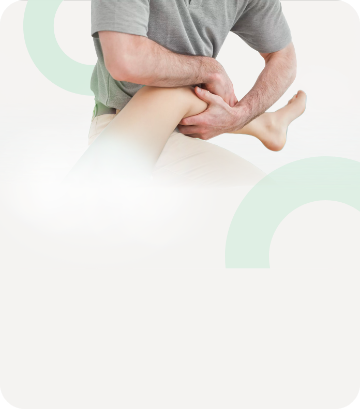
- 4 manual therapy sessions per day.
- Manual therapy
- reduce lumbar tension at the L4 level with ligamentous articular strain techniques;
- high-velocity low-amplitude manipulation of the mid-lumbar segments;
- strain and counterstrain techniques and soft tissue mobilization at the level of the painful area of the left inguinal canal.
Results
After the first session, the pain decreased by 3-4 points out of 10 and the patient was able to get off the treatment table and walk slowly out of the office. After 4 days of treatment, the symptoms disappeared completely.
While a new x-ray continued to show lumbar scoliosis, the tender areas in L4 and inguinal canal had recovered. Similarly, the urinary and bowel problems disappeared along with the pain while the ROM went back to normal and the Barthel Index showed a score of 95 out of 100. After 5 days, the patient was discharged from the hospital with the recommendation to perform a series of exercises at home, including deep breathing, stretching, core stabilization, Taiji and mechanical education to better perform his work as a farmer.
6 months after discharge, the patient reported by telephone that he no longer had symptoms and had returned to work safely.
Discussion
To successfully treat the patient it was necessary to understand the cause of the pain, for which it was in turn necessary to carefully evaluate all the tests available.
Once the ilioinguinal nerve was identified as the main culprit, the articulatory techniques allowed the myofascial tissue of the lumbar area to be released by “freeing” the space between the psoas major and the transverse lumbar processes. Strain and counterstrain and soft tissue techniques reduced abdominal muscle tension and relaxed the areas where the ilioinguinal and iliohypogastric nerves traverse the transverse abdominal muscle and internal oblique muscle near the left inguinal canal.
Of course, the involvement of other structures cannot be ruled out with certainty. However, this case illustrates how a thorough examination of the patient’s history and current status, together with an anatomically grounded diagnostic process can allow for successful therapy.
Manual therapy, therefore, can be a valid tool in similar cases with symptoms closely related to the anatomy, although more in-depth studies are needed to better understand the exact effect of manual therapy.
The review of Osteopedia
By Marco Chiera
Strengths: accurate description of the patient and above all of the clinical reasoning that led to the diagnosis and therefore to the intervention; the tables and anatomical images reported as a support are useful; good description of the intervention; 6-month follow-up; the educational program provided to the patient for self-management is interesting and fundamental.
Limits: like any case report, it is difficult to generalize the results. Although knowing the anatomy is fundamental and certainly useful in the case of acute and sudden pain such as the one presented in this study, we must avoid a purely structural drift of pain, as we now know that pain is a complex response elaborated by the body to front of signals with a high danger threshold, which can come from any level (physical, chemical, biochemical, psychological, social, environmental).
Are you an osteopath?
Register and enjoy the membership benefits. Create your public profile and publish your studies. It's free!
Register now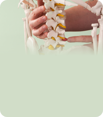
School or training institution?
Register and enjoy the membership benefits. Create your public profile and publish your studies. It's free!
Register now
Do you want to become an osteopath? Are you a student?
Register and enjoy the membership benefits. Create your public profile and publish your studies. It's free!
Register now